{
  "id": 253698,
  "title": "Notes on MRI, Parameter Weighting, and Imaging Planes",
  "url": "/competitions/rsna-miccai-brain-tumor-radiogenomic-classification/discussion/253698",
  "author_name": "",
  "post_date": "2021-07-18T05:35:26.076649800Z",
  "votes": 19,
  "comment_count": 1,
  "views": 0,
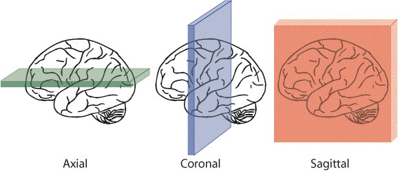
  "content": "<h1>Magnetic resonance imaging (MRI)</h1>\n<h2>Definition</h2>\n<ul>\n<li>MRI is an imaging modality that uses non-ionizing radiation to create useful diagnostic images.</li>\n<li>MRI is used to distinguish pathologic tissue such as a brain tumor or MS lesions from normal tissue.</li>\n<li>In simple terms, an MRI scanner consists of a large, powerful magnet in which the patient lies. A <a href=\"https://radiopaedia.org/articles/radiofrequency-transmitter?lang=us\" target=\"_blank\">radio wave antenna</a> is used to send signals to the body and then a <a href=\"https://radiopaedia.org/articles/radiofrequency-receiver?lang=us\" target=\"_blank\">radiofrequency receiver</a> detects the emitted signals. These returning signals are converted into images by a computer attached to the scanner. Imaging of any part of the body can be obtained in any plane. </li>\n</ul>\n<p>Source: <a href=\"https://radiopaedia.org/articles/mri-2?lang=us\" target=\"_blank\">MRI | Radiology Reference Article | Radiopaedia.org</a></p>\n<h2>Parameter Weighting</h2>\n<p>Following are some of the main parameter weighting techniques being used in MRI:</p>\n<p><img src=\"https://geekymedics.com/wp-content/uploads/2020/04/t1-and-t2-770x287.png\" alt=\"\"></p>\n<h3>T1-weighted</h3>\n<ul>\n<li><strong>ONE</strong> tissue is bright: <strong>fat</strong></li>\n<li>provides the most anatomically-relevant images</li>\n<li>shape of the brain can be clearly seen, and morphological abnormalities are easy to detect.</li>\n<li>fat is depicted in white and water in black.</li>\n<li>grey matter is darker than white matter.</li>\n</ul>\n<h3>T2-weighted</h3>\n<ul>\n<li><strong>TWO</strong> tissues are bright: <strong>fat</strong> and <strong>water</strong> (<strong>WW2</strong> – <strong>W</strong>ater is <strong>W</strong>hite in T<strong>2</strong>)</li>\n<li>Regular T2 MRI's are important for tracking long-term disease progression.</li>\n<li>White matter is darker than grey</li>\n<li>Lesions appear white.</li>\n<li>Suitable for lesion evaluation.</li>\n</ul>\n<h3>T1Gd</h3>\n<ul>\n<li>A Gadolinium (gd)-Enhanced T1-Weighted scan reveals only new lesions. These are areas where the disease is currently active.</li>\n<li>Before the MRI, an injection of gadolinium (gd) is administered. This will distinguish the active lesions from the normal parts of the brain.</li>\n<li>This type of MRI will not show older, inactive lesions.</li>\n</ul>\n<h3>Fluid Attenuated Inversion Recovery (FLAIR)</h3>\n<ul>\n<li>FLAIR is similar to T2, but the fluid is darker or \"suppressed\".</li>\n<li>In T2, the spinal fluid (water) is white and the lesion is also white, so you have to look for the white in the white, which is difficult to understand. <strong>FLAIR</strong> can be roughly thought of as T2, in which the water is also black, making it easier to find the lesion.  </li>\n</ul>\n<h2>Imaging Planes</h2>\n<p>Magnetic resonance imaging (MRI) is partially defined by the plane or direction of the image that is taken. The most important model coordinate system for medical imaging is the <strong>anatomical coordinate system</strong> (also called patient coordinate system). </p>\n<h3>The Three Basic Anatomical Planes:</h3>\n<p>This coordinate system consists of three planes to describe the standard anatomical position of a human. The basic orientation terms for an MRI of the body taken: From the side would be a <strong>sagittal</strong> plane; from the front would be a <strong>coronal</strong> plane, and from the top down would be a <strong>transverse</strong> or <strong>axial</strong> plane.<br>\n<img src=\"https://users.fmrib.ox.ac.uk/~stuart/thesis/chapter_3/image3_5.gif\" alt=\"\"><br>\nSource: <a href=\"https://users.fmrib.ox.ac.uk/~stuart/thesis/chapter_3/image3_5.gif\" target=\"_blank\">https://users.fmrib.ox.ac.uk/~stuart/thesis/chapter_3/section3_2.html</a></p>\n<h4>Sagittal plane</h4>\n<ul>\n<li>Also known as <strong>median plane</strong>.</li>\n<li>It is a y-z plane, perpendicular to the ground, which separates left from right.</li>\n<li>The mid-sagittal plane is the specific sagittal plane that is exactly in the middle of the body.</li>\n<li>A sagittal MRI looks at the brain from the side in a series of images starting at one ear and moving to the other.</li>\n</ul>\n<h4>Coronal plane</h4>\n<ul>\n<li>Also known as <strong>frontal plane</strong>.</li>\n<li>It is an x-z plane, perpendicular to the ground, which (in humans) separates the anterior from the posterior, the front from the back, the ventral from the dorsal.</li>\n<li>A coronal MRI looks at the brain from behind in a series of images starting at the back of the head and moving to the face.</li>\n</ul>\n<h4>Axial plane</h4>\n<ul>\n<li>Also known as <strong>transverse plane</strong>.</li>\n<li>It is an x-y-z plane, parallel to the ground, which (in humans) separates the superior from the inferior, or put another way, the head from the feet.</li>\n<li>An axial MRI looks at the brain from below in a series of images starting at the chin and moving to the top of the head.</li>\n</ul>\n<h2>Sources</h2>\n<ul>\n<li><a href=\"https://my-ms.org/mri_basics.htm\" target=\"_blank\">MRI Basics (my-ms.org)</a></li>\n<li><a href=\"https://www.kaggle.com/c/rsna-miccai-brain-tumor-radiogenomic-classification/discussion/252843\" target=\"_blank\">Differences in MRI imaging methods - MRIの撮影方法の違いについて | Kaggle</a></li>\n<li><a href=\"https://geekymedics.com/the-basics-of-mri-interpretation/\" target=\"_blank\">The Basics of MRI Interpretation | Radiology | Geeky Medics</a></li>\n</ul>",
  "messages": [
    {
      "id": "1391811",
      "postDate": "07/18/2021 05:35:26",
      "content": "<h1>Magnetic resonance imaging (MRI)</h1>\n<h2>Definition</h2>\n<ul>\n<li>MRI is an imaging modality that uses non-ionizing radiation to create useful diagnostic images.</li>\n<li>MRI is used to distinguish pathologic tissue such as a brain tumor or MS lesions from normal tissue.</li>\n<li>In simple terms, an MRI scanner consists of a large, powerful magnet in which the patient lies. A <a href=\"https://radiopaedia.org/articles/radiofrequency-transmitter?lang=us\" target=\"_blank\">radio wave antenna</a> is used to send signals to the body and then a <a href=\"https://radiopaedia.org/articles/radiofrequency-receiver?lang=us\" target=\"_blank\">radiofrequency receiver</a> detects the emitted signals. These returning signals are converted into images by a computer attached to the scanner. Imaging of any part of the body can be obtained in any plane. </li>\n</ul>\n<p>Source: <a href=\"https://radiopaedia.org/articles/mri-2?lang=us\" target=\"_blank\">MRI | Radiology Reference Article | Radiopaedia.org</a></p>\n<h2>Parameter Weighting</h2>\n<p>Following are some of the main parameter weighting techniques being used in MRI:</p>\n<p><img src=\"https://geekymedics.com/wp-content/uploads/2020/04/t1-and-t2-770x287.png\" alt=\"\"></p>\n<h3>T1-weighted</h3>\n<ul>\n<li><strong>ONE</strong> tissue is bright: <strong>fat</strong></li>\n<li>provides the most anatomically-relevant images</li>\n<li>shape of the brain can be clearly seen, and morphological abnormalities are easy to detect.</li>\n<li>fat is depicted in white and water in black.</li>\n<li>grey matter is darker than white matter.</li>\n</ul>\n<h3>T2-weighted</h3>\n<ul>\n<li><strong>TWO</strong> tissues are bright: <strong>fat</strong> and <strong>water</strong> (<strong>WW2</strong> – <strong>W</strong>ater is <strong>W</strong>hite in T<strong>2</strong>)</li>\n<li>Regular T2 MRI's are important for tracking long-term disease progression.</li>\n<li>White matter is darker than grey</li>\n<li>Lesions appear white.</li>\n<li>Suitable for lesion evaluation.</li>\n</ul>\n<h3>T1Gd</h3>\n<ul>\n<li>A Gadolinium (gd)-Enhanced T1-Weighted scan reveals only new lesions. These are areas where the disease is currently active.</li>\n<li>Before the MRI, an injection of gadolinium (gd) is administered. This will distinguish the active lesions from the normal parts of the brain.</li>\n<li>This type of MRI will not show older, inactive lesions.</li>\n</ul>\n<h3>Fluid Attenuated Inversion Recovery (FLAIR)</h3>\n<ul>\n<li>FLAIR is similar to T2, but the fluid is darker or \"suppressed\".</li>\n<li>In T2, the spinal fluid (water) is white and the lesion is also white, so you have to look for the white in the white, which is difficult to understand. <strong>FLAIR</strong> can be roughly thought of as T2, in which the water is also black, making it easier to find the lesion.  </li>\n</ul>\n<h2>Imaging Planes</h2>\n<p>Magnetic resonance imaging (MRI) is partially defined by the plane or direction of the image that is taken. The most important model coordinate system for medical imaging is the <strong>anatomical coordinate system</strong> (also called patient coordinate system). </p>\n<h3>The Three Basic Anatomical Planes:</h3>\n<p>This coordinate system consists of three planes to describe the standard anatomical position of a human. The basic orientation terms for an MRI of the body taken: From the side would be a <strong>sagittal</strong> plane; from the front would be a <strong>coronal</strong> plane, and from the top down would be a <strong>transverse</strong> or <strong>axial</strong> plane.<br>\n<img src=\"https://users.fmrib.ox.ac.uk/~stuart/thesis/chapter_3/image3_5.gif\" alt=\"\"><br>\nSource: <a href=\"https://users.fmrib.ox.ac.uk/~stuart/thesis/chapter_3/image3_5.gif\" target=\"_blank\">https://users.fmrib.ox.ac.uk/~stuart/thesis/chapter_3/section3_2.html</a></p>\n<h4>Sagittal plane</h4>\n<ul>\n<li>Also known as <strong>median plane</strong>.</li>\n<li>It is a y-z plane, perpendicular to the ground, which separates left from right.</li>\n<li>The mid-sagittal plane is the specific sagittal plane that is exactly in the middle of the body.</li>\n<li>A sagittal MRI looks at the brain from the side in a series of images starting at one ear and moving to the other.</li>\n</ul>\n<h4>Coronal plane</h4>\n<ul>\n<li>Also known as <strong>frontal plane</strong>.</li>\n<li>It is an x-z plane, perpendicular to the ground, which (in humans) separates the anterior from the posterior, the front from the back, the ventral from the dorsal.</li>\n<li>A coronal MRI looks at the brain from behind in a series of images starting at the back of the head and moving to the face.</li>\n</ul>\n<h4>Axial plane</h4>\n<ul>\n<li>Also known as <strong>transverse plane</strong>.</li>\n<li>It is an x-y-z plane, parallel to the ground, which (in humans) separates the superior from the inferior, or put another way, the head from the feet.</li>\n<li>An axial MRI looks at the brain from below in a series of images starting at the chin and moving to the top of the head.</li>\n</ul>\n<h2>Sources</h2>\n<ul>\n<li><a href=\"https://my-ms.org/mri_basics.htm\" target=\"_blank\">MRI Basics (my-ms.org)</a></li>\n<li><a href=\"https://www.kaggle.com/c/rsna-miccai-brain-tumor-radiogenomic-classification/discussion/252843\" target=\"_blank\">Differences in MRI imaging methods - MRIの撮影方法の違いについて | Kaggle</a></li>\n<li><a href=\"https://geekymedics.com/the-basics-of-mri-interpretation/\" target=\"_blank\">The Basics of MRI Interpretation | Radiology | Geeky Medics</a></li>\n</ul>",
      "rawMarkdown": "# Magnetic resonance imaging (MRI)\n\n## Definition\n- MRI is an imaging modality that uses non-ionizing radiation to create useful diagnostic images.\n- MRI is used to distinguish pathologic tissue such as a brain tumor or MS lesions from normal tissue.\n- In simple terms, an MRI scanner consists of a large, powerful magnet in which the patient lies. A [radio wave antenna](https://radiopaedia.org/articles/radiofrequency-transmitter?lang=us) is used to send signals to the body and then a [radiofrequency receiver](https://radiopaedia.org/articles/radiofrequency-receiver?lang=us) detects the emitted signals. These returning signals are converted into images by a computer attached to the scanner. Imaging of any part of the body can be obtained in any plane. \n\nSource: [MRI | Radiology Reference Article | Radiopaedia.org](https://radiopaedia.org/articles/mri-2?lang=us)\n\n## Parameter Weighting\nFollowing are some of the main parameter weighting techniques being used in MRI:\n\n![](https://geekymedics.com/wp-content/uploads/2020/04/t1-and-t2-770x287.png)\n\n### T1-weighted\n- **ONE** tissue is bright: **fat**\n- provides the most anatomically-relevant images\n- shape of the brain can be clearly seen, and morphological abnormalities are easy to detect.\n- fat is depicted in white and water in black.\n- grey matter is darker than white matter.\n\n### T2-weighted\n- **TWO** tissues are bright: **fat** and **water** (**WW2** – **W**ater is **W**hite in T**2**)\n- Regular T2 MRI's are important for tracking long-term disease progression.\n- White matter is darker than grey\n- Lesions appear white.\n- Suitable for lesion evaluation.\n\n### T1Gd\n- A Gadolinium (gd)-Enhanced T1-Weighted scan reveals only new lesions. These are areas where the disease is currently active.\n- Before the MRI, an injection of gadolinium (gd) is administered. This will distinguish the active lesions from the normal parts of the brain.\n- This type of MRI will not show older, inactive lesions.\n\n### Fluid Attenuated Inversion Recovery (FLAIR) \n- FLAIR is similar to T2, but the fluid is darker or \"suppressed\".\n- In T2, the spinal fluid (water) is white and the lesion is also white, so you have to look for the white in the white, which is difficult to understand. **FLAIR** can be roughly thought of as T2, in which the water is also black, making it easier to find the lesion.  \n\n## Imaging Planes\nMagnetic resonance imaging (MRI) is partially defined by the plane or direction of the image that is taken. The most important model coordinate system for medical imaging is the **anatomical coordinate system** (also called patient coordinate system). \n\n### The Three Basic Anatomical Planes:\nThis coordinate system consists of three planes to describe the standard anatomical position of a human. The basic orientation terms for an MRI of the body taken: From the side would be a **sagittal** plane; from the front would be a **coronal** plane, and from the top down would be a **transverse** or **axial** plane.\n![](https://users.fmrib.ox.ac.uk/~stuart/thesis/chapter_3/image3_5.gif)\nSource: [https://users.fmrib.ox.ac.uk/~stuart/thesis/chapter_3/section3_2.html](https://users.fmrib.ox.ac.uk/~stuart/thesis/chapter_3/image3_5.gif)\n#### Sagittal plane\n- Also known as **median plane**.\n- It is a y-z plane, perpendicular to the ground, which separates left from right.\n- The mid-sagittal plane is the specific sagittal plane that is exactly in the middle of the body.\n- A sagittal MRI looks at the brain from the side in a series of images starting at one ear and moving to the other.\n\n#### Coronal plane\n- Also known as **frontal plane**.\n- It is an x-z plane, perpendicular to the ground, which (in humans) separates the anterior from the posterior, the front from the back, the ventral from the dorsal.\n- A coronal MRI looks at the brain from behind in a series of images starting at the back of the head and moving to the face.\n\n#### Axial plane\n- Also known as **transverse plane**.\n- It is an x-y-z plane, parallel to the ground, which (in humans) separates the superior from the inferior, or put another way, the head from the feet.\n- An axial MRI looks at the brain from below in a series of images starting at the chin and moving to the top of the head.\n\n## Sources\n- [MRI Basics (my-ms.org)](https://my-ms.org/mri_basics.htm)\n- [Differences in MRI imaging methods - MRIの撮影方法の違いについて | Kaggle](https://www.kaggle.com/c/rsna-miccai-brain-tumor-radiogenomic-classification/discussion/252843)\n- [The Basics of MRI Interpretation | Radiology | Geeky Medics](https://geekymedics.com/the-basics-of-mri-interpretation/)",
      "votes": null
    },
    {
      "id": "1543617",
      "postDate": "10/13/2021 17:25:33",
      "content": "<p>how will your model improve if you decide to utilize mri planes (coronal, sagittal , axial) from dicom metadata , how would you even use in your model?</p>",
      "rawMarkdown": "how will your model improve if you decide to utilize mri planes (coronal, sagittal , axial) from dicom metadata , how would you even use in your model?",
      "votes": null
    }
  ],
  "comments": [
    {
      "id": 1543617,
      "author_name": "aniarya",
      "author_url": "",
      "post_date": "10/13/2021 17:25:33",
      "content": "<p>how will your model improve if you decide to utilize mri planes (coronal, sagittal , axial) from dicom metadata , how would you even use in your model?</p>",
      "votes": null,
      "replies": []
    }
  ],
  "raw_markdown_by_id": {
    "1391811": "# Magnetic resonance imaging (MRI)\n\n## Definition\n- MRI is an imaging modality that uses non-ionizing radiation to create useful diagnostic images.\n- MRI is used to distinguish pathologic tissue such as a brain tumor or MS lesions from normal tissue.\n- In simple terms, an MRI scanner consists of a large, powerful magnet in which the patient lies. A [radio wave antenna](https://radiopaedia.org/articles/radiofrequency-transmitter?lang=us) is used to send signals to the body and then a [radiofrequency receiver](https://radiopaedia.org/articles/radiofrequency-receiver?lang=us) detects the emitted signals. These returning signals are converted into images by a computer attached to the scanner. Imaging of any part of the body can be obtained in any plane. \n\nSource: [MRI | Radiology Reference Article | Radiopaedia.org](https://radiopaedia.org/articles/mri-2?lang=us)\n\n## Parameter Weighting\nFollowing are some of the main parameter weighting techniques being used in MRI:\n\n![](https://geekymedics.com/wp-content/uploads/2020/04/t1-and-t2-770x287.png)\n\n### T1-weighted\n- **ONE** tissue is bright: **fat**\n- provides the most anatomically-relevant images\n- shape of the brain can be clearly seen, and morphological abnormalities are easy to detect.\n- fat is depicted in white and water in black.\n- grey matter is darker than white matter.\n\n### T2-weighted\n- **TWO** tissues are bright: **fat** and **water** (**WW2** – **W**ater is **W**hite in T**2**)\n- Regular T2 MRI's are important for tracking long-term disease progression.\n- White matter is darker than grey\n- Lesions appear white.\n- Suitable for lesion evaluation.\n\n### T1Gd\n- A Gadolinium (gd)-Enhanced T1-Weighted scan reveals only new lesions. These are areas where the disease is currently active.\n- Before the MRI, an injection of gadolinium (gd) is administered. This will distinguish the active lesions from the normal parts of the brain.\n- This type of MRI will not show older, inactive lesions.\n\n### Fluid Attenuated Inversion Recovery (FLAIR) \n- FLAIR is similar to T2, but the fluid is darker or \"suppressed\".\n- In T2, the spinal fluid (water) is white and the lesion is also white, so you have to look for the white in the white, which is difficult to understand. **FLAIR** can be roughly thought of as T2, in which the water is also black, making it easier to find the lesion.  \n\n## Imaging Planes\nMagnetic resonance imaging (MRI) is partially defined by the plane or direction of the image that is taken. The most important model coordinate system for medical imaging is the **anatomical coordinate system** (also called patient coordinate system). \n\n### The Three Basic Anatomical Planes:\nThis coordinate system consists of three planes to describe the standard anatomical position of a human. The basic orientation terms for an MRI of the body taken: From the side would be a **sagittal** plane; from the front would be a **coronal** plane, and from the top down would be a **transverse** or **axial** plane.\n![](https://users.fmrib.ox.ac.uk/~stuart/thesis/chapter_3/image3_5.gif)\nSource: [https://users.fmrib.ox.ac.uk/~stuart/thesis/chapter_3/section3_2.html](https://users.fmrib.ox.ac.uk/~stuart/thesis/chapter_3/image3_5.gif)\n#### Sagittal plane\n- Also known as **median plane**.\n- It is a y-z plane, perpendicular to the ground, which separates left from right.\n- The mid-sagittal plane is the specific sagittal plane that is exactly in the middle of the body.\n- A sagittal MRI looks at the brain from the side in a series of images starting at one ear and moving to the other.\n\n#### Coronal plane\n- Also known as **frontal plane**.\n- It is an x-z plane, perpendicular to the ground, which (in humans) separates the anterior from the posterior, the front from the back, the ventral from the dorsal.\n- A coronal MRI looks at the brain from behind in a series of images starting at the back of the head and moving to the face.\n\n#### Axial plane\n- Also known as **transverse plane**.\n- It is an x-y-z plane, parallel to the ground, which (in humans) separates the superior from the inferior, or put another way, the head from the feet.\n- An axial MRI looks at the brain from below in a series of images starting at the chin and moving to the top of the head.\n\n## Sources\n- [MRI Basics (my-ms.org)](https://my-ms.org/mri_basics.htm)\n- [Differences in MRI imaging methods - MRIの撮影方法の違いについて | Kaggle](https://www.kaggle.com/c/rsna-miccai-brain-tumor-radiogenomic-classification/discussion/252843)\n- [The Basics of MRI Interpretation | Radiology | Geeky Medics](https://geekymedics.com/the-basics-of-mri-interpretation/)",
    "1543617": "how will your model improve if you decide to utilize mri planes (coronal, sagittal , axial) from dicom metadata , how would you even use in your model?"
  },
  "source": "meta"
}